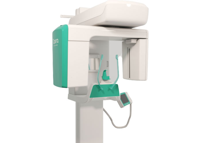
New Life Radiology introduces the most advanced CBCT – CONE BEAM COMPUTED TOMOGRAPHY – technology, the technic of biomedical imaging for the acquisition of volumetric images of the dental arch. CBCT Sistem represents a valid support for the realization of implantology interventions, general/maxillo-facial surgery, periodontics, endodontics and TMJ.
CMOS Flat panel sensor 13 x 13 cm Active Area with 100nm pixel size
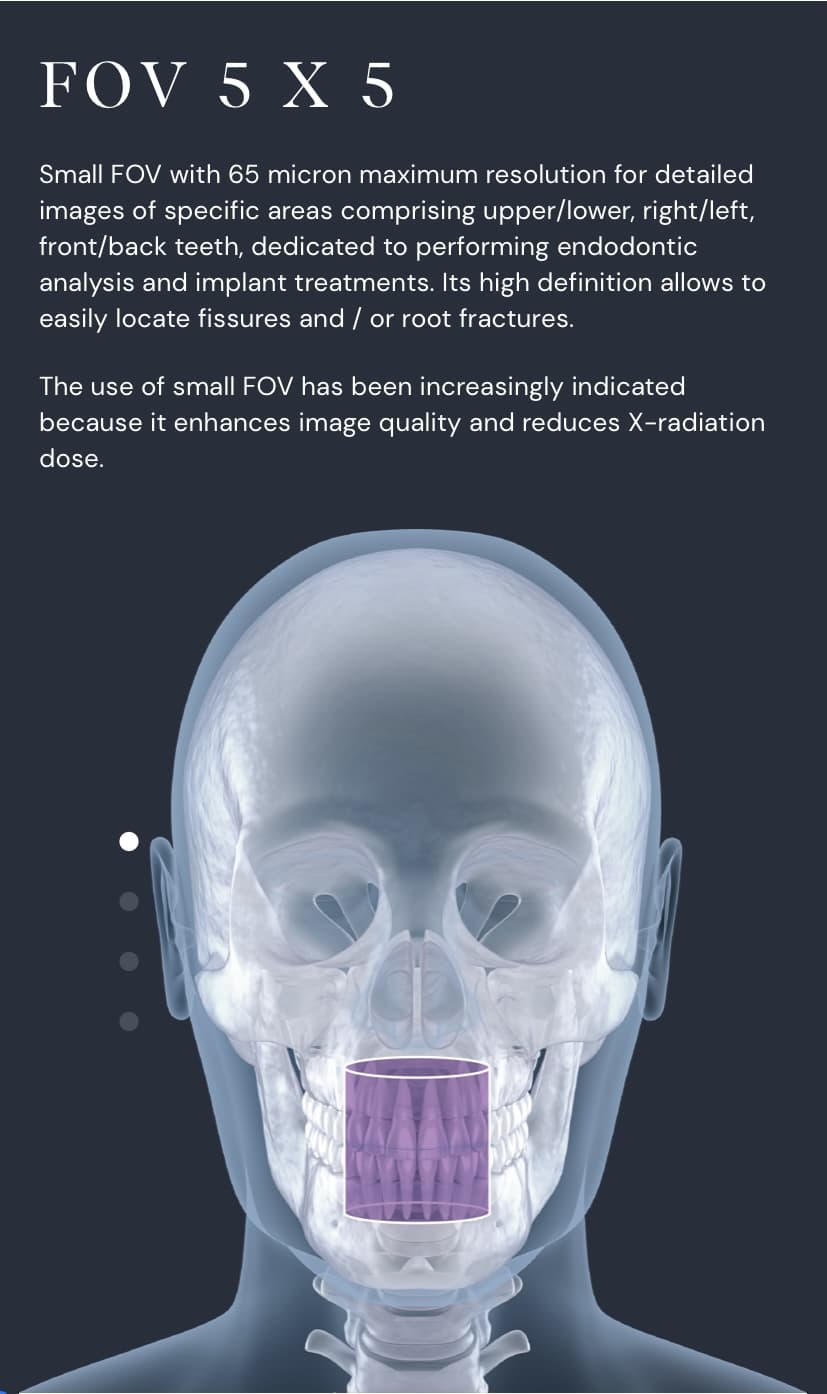
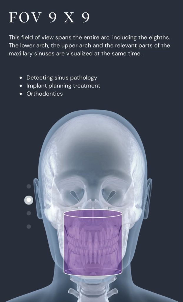
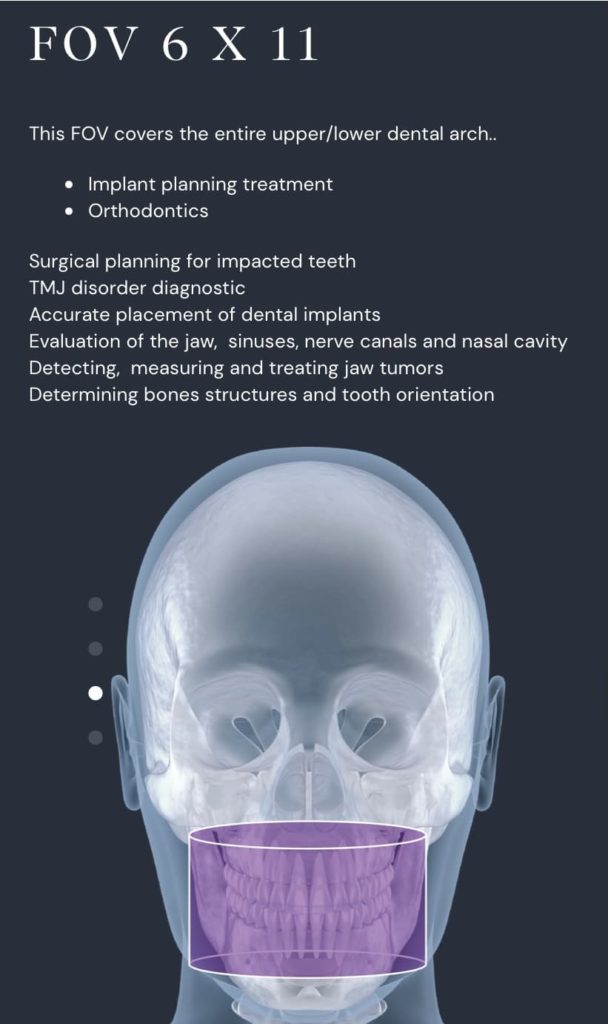
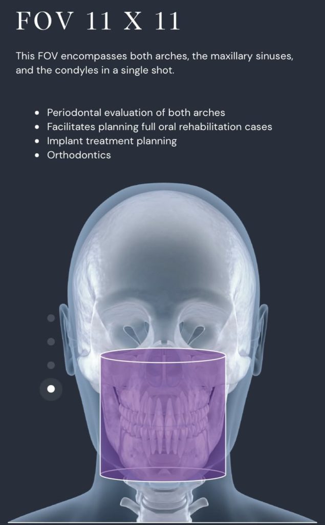
Real FOV available 9×9, 11×11, MULTIFOV (9×9, 11×11, 6×11, 5×5) (not stitching)
Recently developed geometrical calibration
Post-process function for 2D images obtained with the 3D sensor
Same sensor for 2D and 3D images: 2 in 1 solution
An efficient two-in-one solution to obtain 13 x 30 cm 2D images and 9 x 9 (or 11 x11 or MULTIFOV 9×9, 11×11, 6×11, 5×5 cm) Volumetric Images in just 10 seconds.
A CMOS flat panel sensor with 13 x 13 cm active area, 100 micron resolution and capture capacity of 300 frames: with those special features, the acquisition of an ideal images database for volumetric construction of an image with FOV 9 cm (diameter) x 9 cm (height) is guarantee (or 11×11, or MULTIFOV 9×9, 11×11, 6×11, 5×5, depending on chosen configuration)
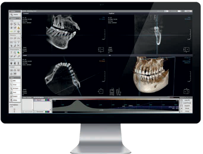
Xelis: Implant planning software
A unique tool to assist you in implant surgery:
Takes transversal cross sections of the dental arch for preliminary implant evaluation and follow up
Clearly indicates the correct position and size of the implants to use
Visualizes with precision the nerve channels and determines the angle for the surgery with greater effectiveness
Xelis has a simple interface to help evaluating numerous clinical pathologies including fracture, included teeth, periodotitis and TMJ.
Xelis advanced implant DBM Xelis Dental Database Basic 3D Toolbar – including Measurements Tools, MPR, Cross Section Advanced Toolbar – Canal Draq / Implant Simulation / Utilities STL export – Save surface CD7DVD7USB export – image export to external storage Batch Print – One click Image Batch Print (Axial, Panoramic, Cross Section) DLB – Dynamic Light Box Image stitching Report – Captured Image Management and Report generation DICOM Print and CD burning Net environment, optional multi user up to 10 users
Xelis basic implant DBM Xelis Dental Database Basic 3D Toolbar – including Measurement Tools, MPR, Cross Section Advanced Toolbar – Canal Draw/Implant Simulation /Utilities Net environment, optional multi user up to 10 users.
Technical specifications for Opera 3D:
3D PANORAMIC Sensor type CMOS Sensor Area 13 x 13 cm Exposure time 14,3/15,0 sec (Child/Adult, Standard PAN) PAN IMAGING PROGRAMS Adult panoramic Child panoramic Option 3 layer focal PAN TMJ closed/open mouth Sinus Sectorial panoramic • Emi Panoramic R • Emi Panoramic L • Low dose Pan (optional) • Ortho panoramic (optional) • Incisors • Bitewing R – Bitewing L – Bitewing r + L (optional) Patient selection : Adult/child, 3 size for all modalities 3D IMAGING Imaging modalities Dentition, TMJ R, TMJ L Field of view 9 x 9 cm (height x diameter) (or 11×11, or MULTIFOV 9×9, 11×11, 6×11, 5×5, depending on chosen configuration)
Detector pixel size 100µm ( 200µm in binning 2×2) Voxel size 121 µm Acquisition rate 2 frames per degree Tube head rotation 230° Dynamic range 14 bit gray level (max 16.384) Number of acquired frames 460 Scan tim/Exp.time 12,2 sec/8,5 sec CEPHALOMETRIC IMAGING Sensor Type DIGITAL FLAT PANEL DR Amorphous Silicon Image Format 24cm x 30cm maximum Exposure type Single shot DR Acquisition time Immediate Setting and exposure time 200ms-500ms CEPH PROGRAMS L/L P/A A/P Carpus projection
X ray generator
Generator type High frequency DC Focal spot 0,5 mm Total filtration > 2,5 mm Aleq @ 70 kV) Leakage radiation According to IEC 60601-2-63: Anodic voltage 61 to 85 kV, step 3 kV Anodic current 4 to 10mA 9 steps Weight 110Kg Dimensions (HxWxD) 2230mm x 1720mm x 1070mm